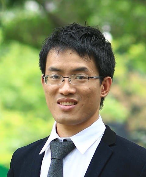
Cheng Jingdong
 Jingdong Cheng, Ph.D.
2005-2009: Shandong University, China, Bachelor.
2006-2007: Xiamen University, China, Exchange student.
2009-2014: Fudan University, China,Ph.D.
2015-2021: Ludwig-Maximilians-Universität München, Germany, PostDoc.
Institutes of Biomedical Sciences, Fudan University, Shanghai 200032
Website:https://chengjdlab.fudan.edu.cn
Email: cheng@fudan.edu.cn
Jingdong is a Principal Investigator of the Institute of Biomedical Sciences (IBS), Fudan University, since 2021. Email: cheng@fudan.edu.cn
Jingdong received his Bachelor degree from Shandong University in 2009. After that, he joined Prof. Yanhui Xu’s lab in IBS, Fudan university, where he received his Ph.D in 2014. During his Ph.D, Jingdong used X-ray crystallography to study the molecular mechanism of DNA methylation mainly mediated by DNMT1/UHRF1/USP7 and DNA demethylation mainly catalyzed by TET family proteins (Cell, 2013; Nature, 2015; JBC, 2013; Nature comm. 2015, 2016; Protein Cell, 2015; et al).
Then, Jingdong moved to Munich, Germany, in 2015 as Postdoc in Prof. Roland Beckmann’s lab. There, Jingdong used cryo-EM single particle analysis to study eukaryotic ribosome biogenesis in yeast, Chaetomium thermophilum and human, including 40S ribosome, 60S ribosome and 39S mitoribosome assembly. By solving plenty intermediates structures, Jingdong revealed the complete detailed assembly process for 40S ribosome start from the nucleolus until the cytoplasm, and the nuclear assembly of the 60S ribosome as well as the late assembly of the 39S mitoribosome. Besides these, Jingdong also did some pioneer work on translational quality control (NSMB, 2017; Nature, 2018; Molecular cell, 2019, 2021; Science 2020; PLoS Biol. 2020; EMBO J, 2021; NAR, 2021; Nature Comm. 2021; et al).
In 2019, Jingdong took the faculty position at Fudan University in Shanghai and established a research laboratory aiming to continue elucidate the mechanisms of human ribosome biogenesis and translational quality control.
Ribosome biogenesis
Translational quantity control
Eukaryotic ribosome formation is a complicated cascade of interdependent biogenesis steps, during which the ribosomal RNA (rRNA) is synthesized, modified, processed and assembled with ca. 80 ribosomal proteins (RPs). Initially, the 18S, 5.8S and 25/28S rRNA are made as a single larger precursor (called 47S pre-rRNA in human), which is co-transcriptionally targeted by snoRNPs at distinct sites, stepwise folded and decorated with ribosomal proteins. These reactions are tightly coordinated by a large set of early assembly factors, which transiently interact with the nascent rRNA and/or r-proteins to form the first pre-ribosomal particles. During the next steps, compaction of the various pre-rRNA domains and remodeling continues until critical pre-rRNA processing steps such as the sequential removal of the external and internal transcribed spacers (5’ ETS, 3’ ETS, ITS1, ITS2) can occur. Further on in the assembly pathway, the pre-ribosomal subunits change location as they move out from the nucleolus into to the nucleoplasm, where intermediate maturation steps are happening. Following a quality control step, the pre-ribosomal particles recruit nuclear export factors and are transported into the cytoplasm, where final maturation and proof-reading generates mature ribosomal subunits. This process already been well studied in yeast or Thermophile, however very little is known for the human cells. In order to close this gap, here, we aim to reveal the assembly mechanisms of the human 40S/60S ribosome by using cryo-EM.
Besides the cytoplasmic 80S ribosome, we also have 55S mitoribosome inside of the mitochondrion, which consist of 28S small subunit and 39S large subunit. The biogenesis of the mitoribosome is completely different compare with 80S ribosome. Evolutionally, its maturation is very similar as its bacterial ancestor, yet it still has many differences. Our previous study, for the first time, revealed the late maturation steps of the human 39S mitoribosome. In our following study, we will further explore the assembly processes both for the 28S and 39S mitoribosome.
Translational quantity control
Under normal condition, ribosome translate the genetic information stored in mRNA into protein starting from AUG start codon until (UAA, UAG or UGA) stop codon. However, if the mRNA is faulty, the ribosome is slowed down or even paused, which in turn will leads to ribosome collision and downstream ribosome quality control pathways. The defective mRNA as well as the not fully translated protein needs to be degraded and the stalled ribosome recycled. Meanwhile, during ribosome biogenesis, defective rRNA and unsuccessful assembled subunits will also trigger ribosome quality control. As a result, the defective ribosomal subunits will be recognized and ubiquitinated, and further will be cleaned up by so far unknown mechanisms. By using structure biology and biochemical methods, we want to uncover the mechanisms in molecular detail.
Cheng J#, Lau B#, La Venuta G, Ameismeier M, Berninghausen O, Hurt E*, Beckmann R*. 90S pre-ribosome transformation into the primordial 40S subunit. Science.2020 Sep 18;369(6510):1470-1476.
Ameismeier M#, Cheng J#, Berninghausen O, Beckmann R. Visualizing late states of human 40S ribosomal subunit maturation. Nature. 2018 June 14; 558 :249-253.
Hu L#, Lu J#, Cheng J#, Rao Q, Li Z, Hou H, Lou Z, Zhang L, Li W, Gong W, Liu M, Sun C, Yin X, Li J, Tan X, Wang P, Wang Y, Fang D, Cui Q, Yang P, He C, Jiang H, Luo C, Xu Y. Structural insight into substrate preference for TET-mediated oxidation. Nature. 2015 Nov 5;527(7576):118-22.
Hu L#, Li Z#, Cheng J#, Rao Q, Gong W, Liu M, Shi YG, Zhu J, Wang P, Xu Y. Crystal structure of TET2-DNA complex: insight into TET-mediated 5mC oxidation. Cell. 2013 Dec 19;155(7):1545-55.